Shoulder Tendon Anatomy Diagram / Anatomy for medical students: Muscles, bones, organs ... / Muscles allow us to move by pulling on bones.. Shoulder muscles and shoulder tendons. This tool is at the same time useful for the training and teaching of the anatomy, but also for experts to illustrate a course or an explanation of pathology to a patient, in particular within the framework of rotator cuff tendon injuries and joint disease. Three bones come together at the shoulder joint. Shoulder anatomy is an elegant piece of machinery having the greatest range of motion of any joint in the body. Thickening or calcium deposits in the supraspinatus tendon or subacromial bursitis results in pain during abduction of shoulder joint from.
The shoulder joint is the connection between the chest and the upper extremity. They are involved in all shoulder motions. The rotator cuff is a group of four muscles and tendons that surround the glenohumeral joint. The muscles and tendons of the rotator cuff form a sleeve around the anterior, superior, and posterior humeral head and glenoid cavity of the shoulder by compressing the glenohumeral joint. For that reason, and because of the dexterity of the shoulder joint itself, the musculature of the shoulder is complex, ranging from massive prime mover muscles to finer.
Anterior shoulder - Health Professions 5061 with Sussman ... from classconnection.s3.amazonaws.com The clavicle (collarbone), the scapula (shoulder blade), and the humerus (upper arm bone) as well as associated muscles, ligaments and tendons. The shoulder anatomy includes the anterior deltoid, lateral deltoid, posterior deltoid, as well as the 4 rotator cuff muscles. Anterior graphic of the shoulder. The human shoulder is made up of three bones: The most important extrinsic soft tissues are the supraspinatus tendon superiorly, infraspinatus posteriorly and subscapularis anteriorly (fig. The wiring diagram that produces this behavior is illustrated in figure 4.4.6. Specifically, the four rotator cuff muscles include the following The shoulder joint is the connection between the chest and the upper extremity.
The bicep has two shoulder tendons:
Each anatomical structure was interactively labeled. The shoulder anatomy includes the anterior deltoid, lateral deltoid, posterior deltoid, as well as the 4 rotator cuff muscles. Ligaments are soft tissue structures that connect bones to bones. The human shoulder is made up of three bones: The shoulder muscles bridge the transitions from the torso into the head/neck area and into the upper extremities of the arms and hands. The subacromial bursa lies on the top portion of the supraspinatus tendon. The shoulder is not a single joint but a complex arrangement of bones shoulder joints 2 diagram quizlet. Related online courses on physioplus. Shoulder muscles and shoulder tendons. Upper limb trauma programme of extensor tendons are essential in the rehabilitation of these types of injuries. A muscle contracts to move bones; An image depicting shoulder anatomy can be seen below. Anterior graphic of the shoulder.
Muscle anatomy dissection 12 photos of the muscle anatomy dissection cat muscle anatomy dissection muscle anatomy dissection human muscles cat muscle anatomy dissection muscle anatomy dissection. The shoulder joint is the connection between the chest and the upper extremity. Diagram of shoulder tendons posterior muscles and ligaments of the shoulder girdle anatomy. The subacromial bursa lies on the top portion of the supraspinatus tendon. Ligaments are soft tissue structures that connect bones to bones.
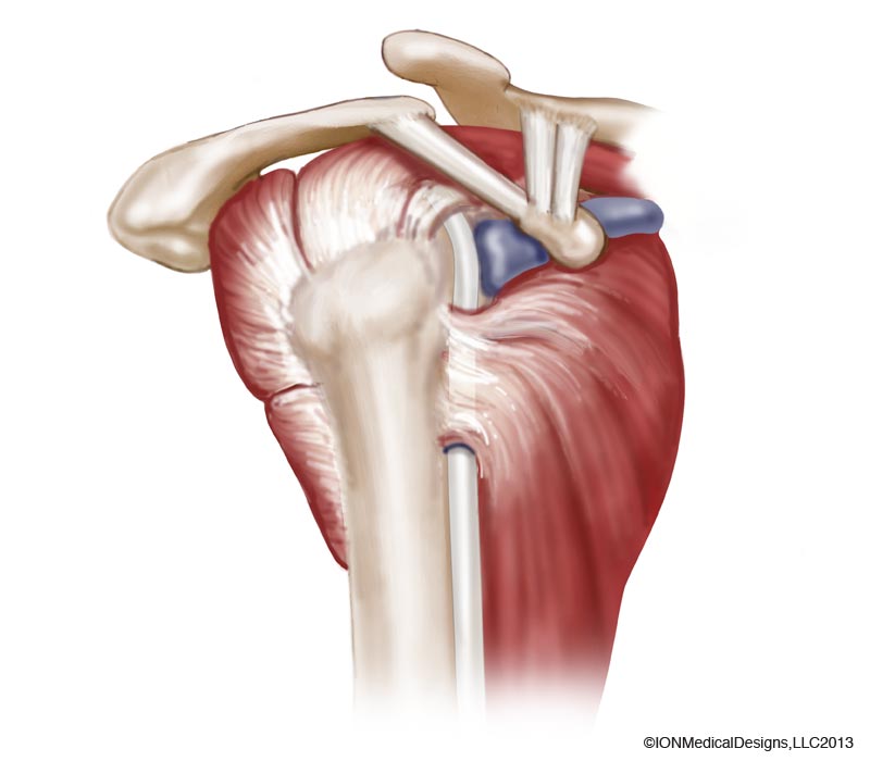
Anatomy of the Shoulder Archives - Joint Preservation Center from josephbermanmd.com They are involved in all shoulder motions. Shoulder muscles and shoulder tendons. Robin smithuis and henk jan van der woude. Diagram of shoulder muscles and tendons / diagram of shoulder tendons shoulder joint anatomyskeletal systemcartilagesligamentsmuscles. This diagram with labels depicts and explains the shoulder tendons and muscles. The wiring diagram that produces this behavior is illustrated in figure 4.4.6. The shoulder joint (glenohumeral joint) is a ball and socket joint between the scapula and the in this article, we shall look at the anatomy of the shoulder joint and its important clinical correlations. The long head and the short head.
Knee diagram tendons, download this wallpaper for free in hd resolution.
Robin smithuis and henk jan van der woude. Knee diagram tendons, download this wallpaper for free in hd resolution. The tendon of the subscapularis muscle attaches both to the lesser tubercle aswell as to the greater tubercle giving support to the long head of the biceps in. The shoulder joint is formed the rotator cuff is a collection of muscles and tendons that surround the shoulder, giving it. Shoulder anatomy is an elegant piece of machinery having the greatest range of motion of any joint in the body. The rotator cuff tendons are a group of four tendons that connect the deepest layer of muscles to the humerus. Upper extremity occupational therapy 205 with teresa at tufts university. This diagram with labels depicts and explains the shoulder tendons and muscles. Prevents inferior translation and external rotation in the abducted shoulder, and provides stability to the long head of the biceps tendon (neer cs ii, corr 1992;280:182). Related online courses on physioplus. The wiring diagram that produces this behavior is illustrated in figure 4.4.6. .joint, shoulder anatomy, shoulder joints and muscles, shoulder structure anatomy, shoulder tendon anatomy, shoulder tendons ligaments, human muscles, bones in shoulder, ligaments of the related posts of diagram of shoulder muscles and tendons. Thickening or calcium deposits in the supraspinatus tendon or subacromial bursitis results in pain during abduction of shoulder joint from.
Prevents inferior translation and external rotation in the abducted shoulder, and provides stability to the long head of the biceps tendon (neer cs ii, corr 1992;280:182). Anatomy of the shoulder part 3 (muscular structures). The tendons are the attachment of the. Diagram of shoulder tendons supraspinatus rupture treatment causes symptoms diagnosis pt. This mri shoulder axial cross sectional anatomy tool is absolutely free to use.
Understanding the Shoulder - Doctor Shoulder from doctorshoulder.co.za For that reason, and because of the dexterity of the shoulder joint itself, the musculature of the shoulder is complex, ranging from massive prime mover muscles to finer. Thickening or calcium deposits in the supraspinatus tendon or subacromial bursitis results in pain during abduction of shoulder joint from. Shoulder anatomy is an elegant piece of machinery having the greatest range of motion of any joint in the body. Specifically, the four rotator cuff muscles include the following Shoulder muscles and shoulder tendons. The subacromial bursa lies on the top portion of the supraspinatus tendon. Draw labelled diagram showing the relations of shoulder joint. The most important extrinsic soft tissues are the supraspinatus tendon superiorly, infraspinatus posteriorly and subscapularis anteriorly (fig.
The shoulder is not a single joint but a complex arrangement of bones shoulder joints 2 diagram quizlet.
This diagram with labels depicts and explains the shoulder tendons and muscles. Muscles of the shoulder anatomy pictures and information. Name the arteries and the nerves that supply shoulder joint. This tool is at the same time useful for the training and teaching of the anatomy, but also for experts to illustrate a course or an explanation of pathology to a patient, in particular within the framework of rotator cuff tendon injuries and joint disease. Normal anatomy, variants and checklist. Specifically, the four rotator cuff muscles include the following The human shoulder is made up of three bones: Robin smithuis and henk jan van der woude. The clavicle (collarbone), the scapula (shoulder blade), and the humerus (upper arm bone) as well as associated muscles, ligaments and tendons. Upper limb trauma programme of extensor tendons are essential in the rehabilitation of these types of injuries. Biceps and triceps tendon rupture. Anatomy of the shoulder part 3 (muscular structures). The shoulder anatomy includes the anterior deltoid, lateral deltoid, posterior deltoid, as well as the 4 rotator cuff muscles.
The bicep has two shoulder tendons: shoulder anatomy diagram. The shoulder joint is the connection between the chest and the upper extremity.


0 Comments